Description
Fusion imaging of anatomic data is proven extremely useful for diagnosis and treatment in radiology, neurology, oncology, and cardiology. It also enables for the localization of tumors and lesions and assists in planning for radiotherapy, biopsy, and surgery with new medical imaging devices.
- Training skills / Applications
- 1. Fusion image generation
- 2. Mapping for biopsy (invasive procedures are not possible with this phantom)
- 3. Surgical planning
- 4. Radiation therapy planning
- 5. Scanning skills in Ultrasound and CT
Specifications
- Anatomy:
- Liver (segmental anatomy, portal and hepatic venous systems, ligamentu, teres and ligamentum venosum)
- Biliary tract (gallbladder, cystic duct, intrahepatic and extrahepatic bile ducts)
- Spleen / kidneys
- Detailed vascular structures (aorta, vena cava, celiac artery and its branches, portal bein and its branches, superior mesenteric vessels, renal vessels, and more)
- Pathology:
Hepatic lesions(cystic and solid)/ Gallbladder and bile duct stones / Pancreatic tumors(one invading the portal bein)/ Splenic lesions / Both kidney lesions / Left adrenal tumor
- Dimensions:
- Size: W29 x D19 x H31 cm
- Weight: 12 kg
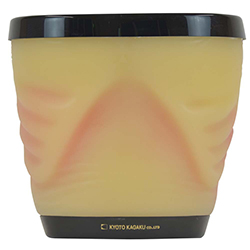
Photos
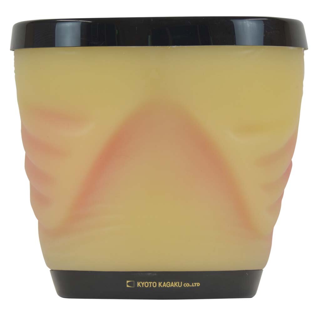
Model
- 41952-000 - Dual Modality Human Abdomen Phantom (CT, Ultrasound) - US-22

